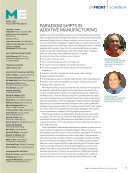
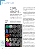
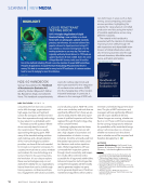
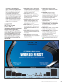
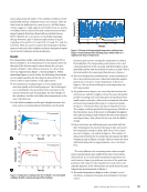
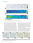
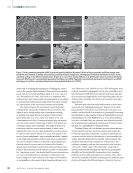
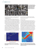
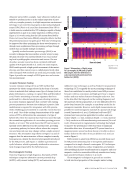
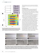
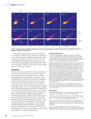
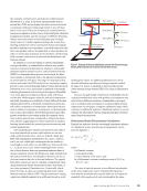
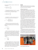
NEW HIP-CT SCANNING TECHNIQUE USES WORLD’S BRIGHTEST X-RAYS Imaging intact human organs from the organ to the cellular scale in three dimensions (3D) has long been a goal of biomedical imaging. To meet this challenge, researchers have recently developed a new technique called hierarchical phase-contrast tomography (HiP-CT), an X-ray phase propagation technique using the European Synchrotron Radiation Facility (ESRF)’s Extremely Brilliant Source (EBS). In November 2021, the authors published a paper in Nature Methods describing how they used the spatial coherence of the ESRF-EBS to perform nondestructive, 3D scans with hierarchi- cally increasing resolution in human organs. HiP-CT provided a structural overview of each whole organ followed by multiple higher-resolution volumes of interest, capturing organotypic functional units and certain individual specialized cells within intact human organs. The technique is providing fresh insights into how COVID-19 damages and reshapes the blood vessels of the lungs. And while its long-term promise is hard to define, because nothing quite like HiP-CT has ever existed before, researchers are excited by its potential. Some 40 different research groups have contacted the team to learn more about the technique. The HiP-CT technique got its start as two German pathologists—Danny Jonigk, a thoracic diseases pathologist at Hannover Medical School, and Maximilian Ackermann, a pathologist at University Medical Center Mainzraced—aimed to track the SARS-CoV-2 virus’s effects across the human body. Medical X-rays such as CT scans can provide a view of an entire organ, but they aren’t high resolution enough. Biopsies can let scientists study tissue samples under a microscope, but the resulting images are only small bits of a whole organ and can’t show how COVID-19 progresses across an entire lung, and the team’s resin technique required dissolving tissue, which destroys the sample and limits further study. Jonigk and Ackermann needed the unprece- dented: a series of X-rays, all done on the same organ, that would let researchers zoom into portions of the organ down to the cellular scale. In March 2020, the German duo reached out to Peter Lee, a materials scientist and chair of emerging technolo- gies at University College London. Lee’s specialty is studying biological materials with powerful X-rays and he quickly realized the potential of the ESRF. The ESRF is located in the northwestern corner of Grenoble, France. The facility is a particle accelerator that makes electrons travel at nearly the speed of light around a half-mile (844 m) circular tunnel. As these electrons speed around the track, powerful magnets along the way bend the particle stream, which causes the electrons to emit the world’s brightest X-rays, which are 100 billion times brighter than the ones used in hospitals. This powerful radiation lets the ESRF examine objects at the scale of micrometers, or even nano- meters. The facility is frequently used to study mate- rials such as alloys and composites, to check the molecular structures of proteins, and even to recon- struct ancient fossils without having to separate rock SCANNER HiP-CT enables 3D imaging of organotypic functional units across intact human organs. Scan QR Code to Watch the Video 8 M A T E R I A L S E V A L U A T I O N • A P R I L 2 0 2 2 CREDIT: REPRINTED WITH PERMISSION UNDER CREATIVE COMMONS LICENSE. C.L. WALSH, P. TAFFOREAU, W.L. WAGNER, ET AL., NATURE METHODS 18, 1532–1541 (2021).

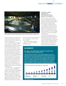
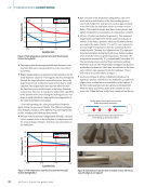
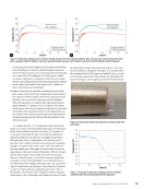
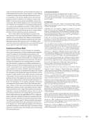
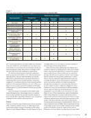
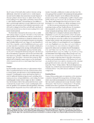
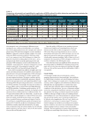
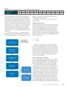
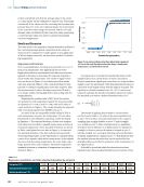
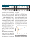
from bone. Ackermann, Jonigk, and Lee wanted to use this huge instrument to perform the world’s most detailed X-ray scans of a human organ. But for all HiP-CT’s promise, there are also considerable challenges. HiP-CT scans generate a huge amount of data— several terabytes per organ. For clini- cians to make real-world use of these scans, the researchers hope to develop a cloud-based interface to navigate them, like Google Maps for the human body. They also need to simplify the process of creating workable 3D models from the scans. Like all CT scanning techniques, HiP-CT works by making many 2D slices of a given object and stacking them together. Much of this process is manual, especially for scans of abnormal or diseased tissues. A major priority for the HiP-CT team is to develop machine-learning techniques to lighten the load. The HiP-CT team is using the ESRF’s newest beam facility, called BM18, to continue scanning human organs. BM18 produces a much bigger X-ray beam, which means scans take less time, and BM18’s X-ray detector can be placed up to 38 m away from the object being scanned, which makes its scans sharper. BM18 also has the space to scan very large objects. Thanks to the new facility, the team’s vision is to scan an entire torso of a human body in one fell swoop by the end of 2023. To read the full paper published in Nature, go to nature.com/articles/ s41592-021-01317-x. CRITICAL AND EMERGING TECHNOLOGIES: AREAS TO WATCH In February 2022, the White House issued an updated list of critical and emerging technologies that the Office of Science and Technology Policy (OSTP), National Science and Technology Council (NSTC), and National Security Council (NSC) have identified as being important for US national security. Last updated in 2020, this list represents a subset of novel, advanced technologies with the potential to chart new pathways in American innovation and strengthen national security. Numerous technology areas have been featured, including advanced engi- neering materials, advanced manufac- turing (including additive manufacturing), advanced nuclear energy technologies, artificial intelligence (including machine learning), autonomous systems and robotics, human-machine interfaces INDUSTRYNEWS | SCANNER DATAFACTS | GLOBAL 3D PRINTING MARKET EXPECTED TO GROW EXPONENTIALLY The global 3D printing market size was valued at US$13.78 billion in 2020 and is expected to expand at a compound annual growth rate (CAGR) of 21.0% from 2021 to 2028. Globally, 2.1 million units of 3D printers were shipped in 2020, and shipments are expected to reach 15.3 million units by 2028. The aggressive R&D in 3D printing (referred to as “additive manufacturing” for industrial applications) and the growing demand for prototyping applications from various industry verticals—particularly healthcare, automotive, aerospace, and defense—are expected to drive market growth. Source: grandviewresearch.com Stereolithography Fused deposition modeling Selective Laser Sintering Direct metal laser sintering Polyjet printing Inkjet printing Electron beam melting Laser metal deposition Digital light processing Laminated object manufacturing Others 2017 1.85 2.21 2018 2019 2020 2021 2022 2023 2024 2025 2026 North America 3D printing market size, by technology, 2017–2028 (USD billion) 2027 2028 Top view of the circular tunnel at the European Synchrotron Radiation Facility in Grenoble, France. A P R I L 2 0 2 2 • M A T E R I A L S E V A L U A T I O N 9 CREATIVE COMMONS ATTRIBUTION-SHARE ALIKE 2.0 FRANCE LICENSE.
ASNT grants non-exclusive, non-transferable license of this material to  .
All rights reserved. © ASNT 2026. To report unauthorized use, contact: customersupport@asnt.org