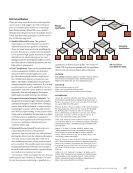
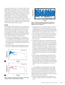
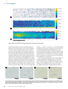
ABSTR ACT Binder jet metallic additive manufacturing (AM) is a popular alternative to powder bed fusion and directed energy deposition because of lower costs, elimination of thermal cycling, and lower energy consumption. However, like other metallic AM processes, binder jetting is prone to defects like porosity, which decreases the adoption of binder-jetted parts. Binder-jetted parts are sometimes infiltrated with a low melting temperature metal to fill pores during sintering however, the infiltration is impacted by the part geometry and infiltration environment, which can cause infill nonuniformity. Furthermore, using an infiltration metal creates a complicated multiphase microstructure substantially different than common wrought materials and alloys. To bring insight to the binder jet/infiltration process toward part qualification and improved part quality, spatially dependent ultrasonic wave speed and attenuation techniques are being applied to help characterize and map porosity in parts made by binder jet AM. In this paper, measurements are conducted on binder-jetted stainless steel and stainless steel infiltrated with bronze samples. X-ray computed tomography (XCT) is used to provide an assessment of porosity. KEYWORDS: ultrasonic testing, X-ray computed tomography, microstructure characterization, porosity Introduction Additive manufacturing (AM) is a direct-write technique in which parts are fabricated in a layered fashion following a computer-aided design. Binder jet is a category of AM in which metal green parts are fabricated at room temperature using a powder bed and an aqueous organic binder. As green binder-jetted parts are weakly bonded through the binder, parts are then sintered to remove the binder and fuse the metal powder. To help improve the density of the part, an infil- tration step is sometimes performed in which a lower melting point metal is wicked into the sintered part (Do et al. 2018). Post-processing heat treatments increase component density however, it remains challenging to remove all porosity in binder-jetted metal alloys (Zhu et al. 2020). As pores are stress concentrators with higher probabilities to initiate fracture and impact mechanical properties, it is important to detect such defects post process (Kurgan 2014). Nondestructive evaluation (NDE) techniques can aid in defect detection as well as char- acterization of process-structure linkages in novel AM pro- cesses. In particular, there is a significant need to develop NDE techniques to characterize final binder-jetted parts (Hassen and Kirka 2018) and identify processing conditions that yield reliable components. X-ray computed tomography (XCT), an NDE technique commonly used to detect discontinuities, has been success- fully applied to detect and characterize pores in AM materials (Thompson et al. 2016). Wilson-Heid et al. (2019) evaluated intentional internal penny-shaped pores with XCT and studied their effects on the tensile properties of laser powder bed fusion fabricated stainless steel 316L (SS316L). Ilogebe et al. (2019) observed the internal pore structure and determined the pore volume fraction of binder-jetted structural amor- phous metals with XCT. Zhu et al. (2020) combined XCT with machine learning to characterize pore evolution of binder- jetted copper after fabrication without any further processing, after sintering, and after hot isostatic pressing. Pores were char- acterized by size, sphericity, and orientation, and the authors noted that pores in sintered samples tend to be smaller, more spherical, and are orientated horizontally due to anisotropic sintering (Zhou et al. 2020). While XCT can be used to char- acterize porosity, the accuracy of the data highly depends on the scanning resolution (the voxel size) pores smaller than the voxel size will not be detected and the minimum detect- able pore size is at least three voxels wide in two dimensions (2D) and at least 27 voxels in volume in three dimensions (3D) ULTRASONIC CHARACTERIZATION OF POROSITY IN COMPONENTS MADE BY BINDER JET ADDITIVE MANUFACTURING BY OLIVIA J. COOK*, NANCY HUANG†, ROBERT L.W. SMITHSON‡, CHRISTOPHER M. KUBE*, ALLISON M. BEESE†§, AND ANDREA P. ARGÜELLES** * Department of Engineering Science and Mechanics, Pennsylvania State University, University Park, PA 16802, USA † Department of Materials Science and Engineering, Pennsylvania State University, University Park, PA 16802,USA ‡ 3M Co., Maplewood, MN 55144, USA § Department of Mechanical Engineering, Pennsylvania State University, University Park, PA 16802, USA ** Department of Engineering Science and Mechanics, Pennsylvania State University, University Park, PA 16802, USA arguelles@psu.edu Materials Evaluation 80 (4): 37–44 https://doi.org/10.32548/2022.me-04266 ©2022 American Society for Nondestructive Testing NDTTECHPAPER | ME A P R I L 2 0 2 2 • M A T E R I A L S E V A L U A T I O N 37

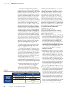
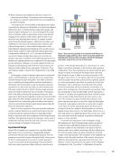
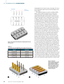
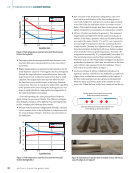
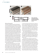
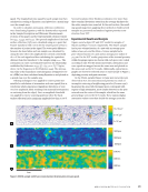
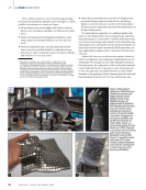
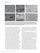
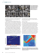
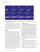
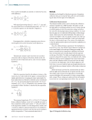
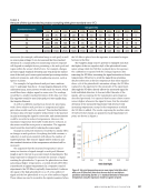
(du Plessis et al. 2018). Trade-offs between resolution, the size of the region of interest, and the time and cost associated with XCT scanning may preclude this technique from widespread implementation for quality control. Ultrasonic testing (UT) has the potential to be used as a porosity detection tool, much like XCT, due to its sensitivity to the elastic properties of the medium through which ultrasonic waves propagate. Compared to XCT, UT is an economical alternative for characterizing the microstructure of binder- jetted parts, enabling inspection of larger part volumes without increased costs. UT has been demonstrated to have measur- able sensitivity to porosity through multiple metrics including wave speed and attenuation (Slotwinski et al. 2014 Daniel et al. 1992). In previous studies, UT has been employed to character- ize the properties of AM parts (Mandache 2019 Honarvar and Varvani-Farahani 2020 Koester et al. 2018). Lopez et al. (2018) studied different nondestructive methods for monitoring wire and arc AM parts and found that UT was a suitable method for both in situ and ex situ defect detection and sizing. Slotwinski et al. (2014) used UT as a method for assessing porosity in CoCr samples made by selective laser melting and found that wave speed correlates well to porosity identified by XCT mea- surement. Ultrasound has also been used to assess the elastic constants of parts made by ultrasonic additive manufacturing (UAM), where UAM-produced parts were found to have up to a 48 reduction in elastic constant values compared to a con- ventionally manufactured sample (Foster et al. 2013). Sotelo et al. (2021) employed immersion ultrasound to measure wave speed and attenuation and characterize the spatially varying properties of hybrid AM samples. With the exception of the authors’ recent paper investigating binder jet printed parts using UT, these methods have not been explored to character- ize binder-jetted components (Huang et al. 2022). In this paper, UT is used to investigate the microstructure and porosity of binder jet AM parts made of stainless steel 316L and stainless steel 316 infiltrated with bronze. Samples are tested in an immersion system in a pulse-echo, normal-incidence con- figuration. Validated ultrasonic methods are merged to generate spatial maps of longitudinal attenuation and wave speed in samples with spatially varying porosity. Ultrasonic maps are then compared to XCT measures of porosity. The results illus- trate the effectiveness of UT in identifying large pore networks in the stainless steel as well as the presence of distributed porosity in the stainless steel infiltrated with bronze. Background While ultrasonic wave speed measurements usually rely on precise knowledge of the sample thickness, Kuo et al. (1990) reported an alternative method that eliminates the need for a priori thickness measurements, and Fei et al. (2001) expanded this method to conduct simultaneous measurement of wave speed and thickness. Figure 1 depicts the test configu- ration, which requires that the sample be tested in pulse-echo mode with a planar reflector directly behind the sample. As shown in Figure 1, two consecutive measurements with and without the sample present are necessary to calculate the time of arrival of the sample front-surface reflection (T1), the sample back-surface reflection (T2), the plate reflection with the sample in the propagation path (TM), and the plate reflection without the sample in the propagation path (TW). From the time of arrival of these reflections, the longitudi- nal wave speed in the sample can be calculated by: (1) c L = W TM) T2 T1) ( +1&c !(T " % # $ water where cwater is the wave speed in the water. The sample thickness (tsample) can then be estimated by: (2) tsample = cL T2 T1) ( 2 which can be used to calculate attenuation. Absolute atten- uation measurements also require consideration of system effects. Various approaches have been proposed, such as those in Lerch et al. (2006), but the equal diffraction method described in Yu et al. (2001) was employed herein. In Yu et al.’s method, a reference sample with near-zero attenuation is measured with identical system settings (such as gain, fre- quency filters, and input voltage) to the test samples. The water paths (WP), or the distance that the wave propagates through water before entering the solid, for the sample and reference, are set to satisfy the equivalent path relation: (3) WP Ref +t Ref cRef c water = WP sample +t sample c sample c water where c is the wave speed, and t is the thickness for the reference (“Ref”) or sample. Reflector Sample Transducer T 1 T 2 T M Reflector Sample Transducer T W Figure 1. Schematic of the experimental configuration for thickness- independent wave speed measurements: (a) the reflections collected when the sample is present (b) the reflection collected after the sample is removed. ME | BINDERJETAM 38 M A T E R I A L S E V A L U A T I O N • A P R I L 2 0 2 2
ASNT grants non-exclusive, non-transferable license of this material to  .
All rights reserved. © ASNT 2026. To report unauthorized use, contact: customersupport@asnt.org